
Posts Tagged: cardiology
Let’s Have a Heart to Heart
Introducing Specialty Veterinary Cardiology Services at Our Primary Care Clinic
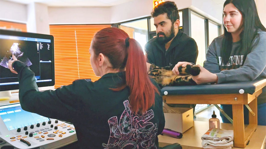
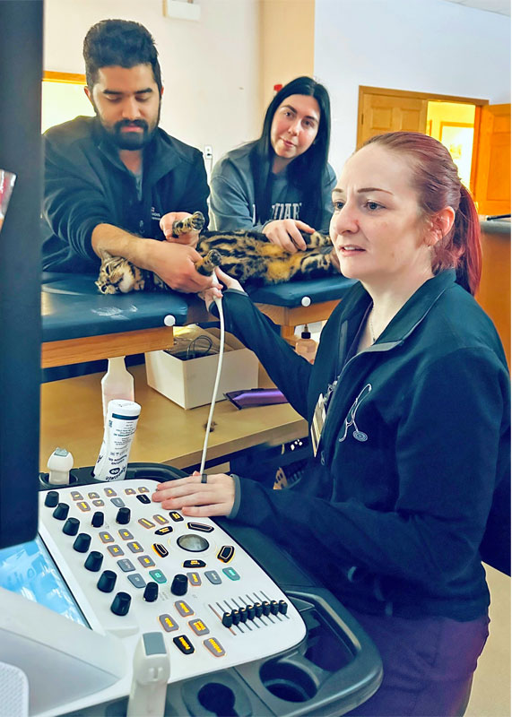
This month, love is in the air, and we’re celebrating the hearts that matter most—the ones inside your beloved pets. We’re excited to announce that our clinic now offers veterinary cardiology specialty services, including echocardiography (heart ultrasound), provided by a board-certified veterinary cardiologist, right here in the clinic you already know and trust.
What Is a Veterinary Cardiologist?
A veterinary cardiologist is a veterinarian with advanced specialty training focused entirely on heart disease. They partner closely with us as your primary care veterinarians to diagnose, monitor, and manage heart conditions—whether your pet is in the early stages of life or enjoying their golden years.
By bringing cardiology services into our primary care setting, we get the best of both worlds: specialist-level expertise paired with the personalized primary care your pet already receives.
An Echocardiogram: A Big Window into Your Pet’s Heart
An echocardiogram is a noninvasive ultrasound that lets us see the heart in motion—how the chambers fill, how the valves open and close, and how effectively the heart pumps blood.
It’s painless, typically does not require sedation, and provides a lot of information that cannot be obtained from X-rays or physical exams alone. It is one of the best ways for diagnosing most forms of heart disease and determining and evaluating what cardiac medications your pet needs. It also gives us clues on anesthetic safety and whether certain sedatives can be used for procedures.
Echocardiography can help diagnose and monitor many cardiac conditions, including:
- Heart murmurs – A heart murmur is an extra whooshing sound heard when the heart is auscultated. An echocardiogram can sort out innocent murmurs (a harmless sound) from true heart disease
- Degenerative (valvular) heart disease – Common in older, small- and medium-breed dogs
- Dilated cardiomyopathy (DCM) – Often seen in large-breed dogs
- Hypertrophic cardiomyopathy (HCM) – The most common heart disease in cats
- Congenital heart defects – Such as PDA or valve abnormalities
- Arrhythmias (abnormal heart rhythms) – When evaluated alongside ECG findings
- Pericardial disease – Fluid around the heart
- Pulmonary hypertension – Increased pressure in the lungs affecting heart function
Why We Love Offering Cardiology Care
From a primary care veterinarian’s perspective, having a specialist work directly within our clinic is truly special and not something we get the advantage of having every day.
A strong heart team: As your primary care veterinarian, we know your pet. Working hand-in-hand with an on-site cardiologist deepens our knowledge of your pet’s health, helps take guesswork out of complex cardiac cases, and allows for stronger decision making recommendations when it comes to things like anesthesia, surgery, and fluid therapy. That teamwork leads to unified treatment plans, better outcomes and fewer complications.
Less stress, more comfort: Vet visits can be stressful for pets and their owners alike. Receiving specialty care in a familiar environment helps pets (and you) feel more at ease.
Faster answers, better continuity: Because cardiology services are available right here, we can remove the specialty bottleneck and move from concern to diagnosis to treatment without unnecessary delays. And with more data on your pet, we can more specifically target treatment plans to your pet’s needs, sooner, and with more confidence.
Expert care with a personal touch: Our cardiologist collaborates closely with our primary care veterinarians to create treatment plans that are effective, realistic, and aligned with your goals for your pet’s health. The streamlined process means you get personalized answers, faster. We love having cardiology right at our fingertips!
When a Cardio Consult Could Help
Your veterinarian may recommend a cardiology evaluation if your pet has:
- A newly detected heart murmur
- Coughing, exercise intolerance, or breathing changes
- Weakness or fainting episodes
- Known heart disease needing monitoring
- Breed-related risk for heart conditions
- Abnormal findings on X-rays or lab work
- A known heart condition and needs an evaluation before an upcoming anesthetic or surgical procedure
You love your pet, and we do too! And now, advanced heart care doesn’t have to mean leaving the clinic you trust. With specialized veterinary cardiology services and primary care all under one roof, your pet can receive expert cardiac care in a place that already feels like home. Don’t hesitate to ask us if a cardiology consult is right for your pet!
— Amanda Hampton, DVM (she/her/hers)
Four Heart Abnormalities Add up to Tetralogy of Fallot

A puppy born with a rare heart condition—Tetralogy of Fallot—got a second chance at life, thanks to a collaboration involving a visiting veterinary heart surgeon and the cardiology and surgery teams at the University of Illinois Veterinary Teaching Hospital.
Odyssey, an Australian shepherd, was only three months old and newly placed in a foster home when he started showing signs of a problem. After exercising, he began breathing heavily and had trouble standing up on his own. He would lean up against the fence just to stay standing and was having uncontrollable tremors. (article continues below)
Cardiology Services at Medical District Vet Clinic
Since November 2025, boarded cardiologists from the University of Illinois Veterinary Teaching Hospital have offered appointments on a part-time basis at the Medical District Veterinary Clinic in Chicago. Services offered include pre-anesthetic echocardiograms, Holter monitoring, management of stable cardiac disease, rechecks, and more. Please call 312-226-2588 to schedule a cardiology appointment for your pet.
Patients with emergent or critical cardiac disease should seek immediate attention and should not wait to see a cardiologist at the clinic.
Dr. Todd Sumerfield, a veterinarian pursuing specialization in veterinary cardiology at the College of Veterinary Medicine, explains how the Illinois team diagnosed Odyssey’s condition and arranged for his surgical intervention—a modified Blalock-Taussig shunt—that had never before been performed at the university hospital.
Diagnosing Tetralogy of Fallot
Noticing Odyssey’s post-exercise behaviors, his foster owners took him to their veterinarian, who identified a loud heart murmur and referred the case to the cardiology team at Illinois in August 2023.
“An echocardiogram was performed, as well as a packed cell volume,” Dr. Sumerfield says.
An echocardiogram allows doctors to visualize the heart as it beats within the patient. In Odyssey’s case, the echocardiogram identified all the defects associated with a condition known as Tetralogy of Fallot.
Packed cell volume measures the amount of red blood cells within the blood of the patient. When an animal has a heart condition leading to decreased oxygen in the blood, as in Tetralogy of Fallot, the body produces more red blood cells in an attempt to deliver more oxygen to the body’s tissues. Although the echocardiographic signs of Odyssey’s condition were severe, his packed cell volume was within the normal range, at 44%.
Dr. Sumerfield points out this normal finding “underlines that Odyssey was doing better than most dogs with this condition at the time of his diagnosis.”
What Is Tetralogy of Fallot?
“Tetralogy of Fallot is an uncommon, congenital heart disease,” Dr. Sumerfield says. “It occurs when the patient has four (thus ‘tetralogy’) concurrent abnormalities.”
To understand these abnormalities, it helps to recall how blood flows through the body. Blood is pumped from the heart’s right atrium to its right ventricle and then, via the pulmonary artery, to the lungs. After receiving oxygen from the lungs, the blood then travels back to the heart into the left atrium. From there it is pumped to the left ventricle, which pushes the blood into the large blood vessel called the aorta and on to the rest of the body.
After delivering its oxygen to tissues and organs, the deoxygenated blood travels back to the right atrium, where the cycle begins again.
In a Tetralogy of Fallot case, the animal is born with a narrowing of the pulmonary valve, which controls blood flow from the right side of the heart to the lungs. The animal also has a ventricular septal defect, which is an inappropriate opening in the septum, or wall, that separates the left and right ventricles. Additionally, the aorta, instead of being positioned over the left ventricle, sits over the left and right ventricular outflow tracts. In that location, the aorta accepts blood from both the left and right ventricles, sending a mixture of oxygenated and deoxygenated blood to the body’s tissues. Lastly, the right ventricle of the heart is abnormally thickened.
Heart Defects Lead to Low Oxygen
Dr. Sumerfield continues, “The narrowing of the pulmonary valve obstructs the outflow of the blood to the lungs. As a consequence, the right ventricle must pump harder to force the same volume of blood through a narrowed orifice.” Because the right ventricle functions just like any other muscle, as it “works out” harder, the muscle grows and thickens.
The ventricular septal defect means deoxygenated blood found in the right ventricle can flow into the left ventricle, where it is pushed into the body without having passed by the lungs to take on oxygen.
All four abnormalities are interrelated and contribute to the problem of low oxygen levels in circulating blood. Abnormally low circulating oxygen led to Odyssey’s exercise intolerance: he was unable to oxygenate his body properly while playing in the yard, making him easily tired.
In response to low oxygen in the body, the bone marrow receives instructions to produce more red blood cells. “When there are inappropriately high numbers of red blood cells in circulation, the blood can become more viscous, or thick. This change, in turn, can impair blood flow to the brain and lead to neurologic signs and even seizures.” Or, in Odyssey’s case, tremors.
Surgery: Modified Blalock-Taussig Shunt
Once Odyssey had a diagnosis, the next step was to determine a treatment approach. It is not typical to treat a patient with Tetralogy of Fallot with medications until there is evidence of heart failure. Phlebotomy (physical reduction of the number of red blood cells) and balloon valvuloplasty (a minimally invasive procedure to widen the pulmonary valve) were considered but both approaches had limitations.
Another option was surgical intervention using the modified Blalock-Taussig shunt. “This surgery involves redirecting mixed arterial blood flow back to the lungs for another pass at oxygenating the red blood cells,” explains Dr. Sumerfield. “The procedure is performed under general anesthesia via a left thoracotomy. A synthetic tube is placed to allow arterial blood to flow to the lungs. The goals of the procedure would be to improve forward flow through the lungs and have a higher percentage of oxygenated blood circulating at any one time.”
A shunt can be thought of as an alternate path, or a turn in the road. It provides the blood with somewhere else to go. Shunts that arise naturally may be present at birth or may be “acquired” by the body after certain stressors. However, in this surgery, an artificial shunt is placed to redirect blood from where it was going into the body (with no oxygen supply) back to the pulmonary artery (which feeds into the lungs) so it can receive its oxygen load.
Odyssey Still Going Strong
Although Odyssey’s clinical signs were not severe, they were getting progressively worse. After his fosters decided to pursue the modified Blalock-Taussig shunt surgery, the University of Illinois cardiology service conferred with surgeons across the country, and Dr. Brian Sutherland, a veterinary cardiothoracic surgeon from the University of Georgia, agreed to perform the surgery. The modified Blalock-Taussig shunt surgery was performed at the Veterinary Teaching Hospital in September 2024, when Odyssey was 1 year, 4 months old. The surgery was successful and Odyssey recovered well.
After two days in the hospital, Odyssey was sent home with pain relievers, sedation, and a strict exercise restriction for two weeks. He was also given six months of blood thinners to decrease the risk of a clot forming in the newly implanted synthetic shunt.
“If left untreated, most dogs with Tetralogy of Fallot will die before reaching one year of age,” says Dr. Sumerfield. “There is limited information regarding life expectancy in dogs who have had the modified Blalock-Taussig shunt procedure, but one journal article found that patients lived for an average of 7 years after the procedure.”
Odyssey is currently more than two years old and continuing to improve. His ability to exercise has improved significantly and his tremors have resolved. Recent rechecks have shown that Odyssey’s shunt is still working.
His fosters have formally adopted Odyssey and are hoping Odyssey lives a long and happy life.
Dilated Cardiomyopathy: The Big Heart Disease

This one’s for all the gentle giants, dashing Danes, and persnickety pinschers.
Owners frequently tell others about their dog’s “big heart.” But what if that is not a reference to the dog’s lovable personality? Boarded cardiologists at the University of Illinois College of Veterinary Medicine explain dilated cardiomyopathy, a situation where a “big heart” is not a good thing. (article continues below)
Cardiology Services at Medical District Vet Clinic
Since November 2025, boarded cardiologists from the University of Illinois Veterinary Teaching Hospital have offered appointments on a part-time basis at the Medical District Veterinary Clinic in Chicago. Services offered include pre-anesthetic echocardiograms, Holter monitoring, management of stable cardiac disease, rechecks, and more. Please call 312-226-2588 to schedule a cardiology appointment for your pet.
Patients with emergent or critical cardiac disease should seek immediate attention and should not wait to see a cardiologist at the clinic.
Dilated cardiomyopathy (DCM) affects the muscles of the heart so that they no longer contract forcefully enough to adequately pump blood. This results in an enlargement of the heart, which in turn further impairs the heart’s ability to contract. As this negative cycle continues, the patient becomes increasingly likely to develop congestive heart failure. If congestive heart failure develops, patients will require daily medication and frequent veterinary appointments to monitor their disease.
Who Gets DCM?
Large breed dogs, such as Doberman pinschers, great Danes, and boxers, that are middle-aged or older are predisposed to developing DCM. It may be advisable to screen dogs for the disease if they have a family history of DCM or a predisposition due to their breed.
Factors other than breed may play a role in dogs developing DCM. Over the past few decades, veterinary researchers have published data supporting a link between certain diets and DCM.
These diets are often labeled “grain-free” since they use peas, lentils, other legumes, and potatoes as the main ingredient. It is recommended that dog owners avoid feeding “grain-free” and other diets containing legumes.
What Does DCM Look Like?
DCM is different from other cardiac diseases because it may be present for a long time before the patient shows clinical signs. Even though the patient appears healthy, the disease is present and progressing.
Clinical signs can vary widely between patients with DCM. The first symptoms may be exercise intolerance or lethargy. Patients who have progressed to congestive heart failure can suffer from coughing, collapse, and respiratory distress.
Unfortunately, in some dogs, sudden collapse and even death could be the first sign that anything is wrong. For this reason, screening for DCM and monitoring for progression of the disease are vitally important.
Because DCM affects the heart muscle, it can also cause abnormal heart rhythms, called arrhythmias. Arrhythmias originate in the ventricles, the parts of the heart that pump blood to our lungs and throughout our bodies. In DCM, arrhythmias can occur at the same time or even before changes in the shape and musculature of the heart.
Managing DCM
DCM cannot be cured, but the disease can be managed. The goals of therapy are to improve the heart’s ability to contract, improve clinical signs such as being tired and out of breath, and delay onset of congestive heart failure.
Oral medications are used to improve the efficiency of the heart’s contractions and promote forward flow of blood. Patients with congestive heart failure require additional oral medications to decrease fluid accumulation.
The long-term prognosis for canine DCM can be extremely variable, with some patients rapidly progressing to congestive heart failure and requiring therapy while others remain stable for years. Once congestive heart failure develops, however, patients usually have a poor prognosis. Doberman pinschers also seem to deteriorate more rapidly than other breeds.
Hope for the Future
Having an animal with dilated cardiomyopathy can be scary, but researchers are working on a better treatment. The cardiology service at the University of Illinois participated in a study that evaluates the use of a drug called rapamycin in patients with DCM. Called the REPAIR study, this multi-clinic clinical trial sought to determine if rapamycin will reduce cardiac size and improve contractility in dogs with DCM.
Rapamycin treatment has been shown to reverse age-related declines in cardiac function in both laboratory animals and client-owned dogs, without significant adverse effects.
Maple’s Murmur: A Chihuahua Sees a Heart Specialist

Maple’s owners noticed a problem. Their nine-year-old Chihuahua started to have a dry, hacking cough three to four times a day, mainly in the morning. After coughing for 5 to 10 seconds, Maple would make a retching sound, lick her lips, and then return to normal.
Years earlier, Maple’s primary veterinarian had detected a heart murmur. But because Maple was asymptomatic and the murmur was quiet at that time, they had decided to monitor for changes rather than starting medications or performing other diagnostics. (article continues below)
Cardiology Services at Medical District Vet Clinic
Since November 2025, boarded cardiologists from the University of Illinois Veterinary Teaching Hospital have offered appointments on a part-time basis at the Medical District Veterinary Clinic in Chicago. Services offered include pre-anesthetic echocardiograms, Holter monitoring, management of stable cardiac disease, rechecks, and more. Please call 312-226-2588 to schedule an appointment for your pet.
Patients with emergent or critical cardiac disease should seek immediate attention and should not wait to see a cardiologist at the clinic.
Now the murmur could be heard on both sides of the chest with a stethoscope. Maple had also developed a mild tracheal (windpipe) sensitivity, seen as a soft cough when her veterinarian touched her throat. The rest of her physical exam was within normal limits. Given Maple’s breed, age, and history of progressive heart murmur, her veterinarian suspected she had developed chronic valve disease and suggested that Maple see a veterinary cardiologist.
Dr. Todd Sumerfield, a veterinarian pursuing specialization in cardiology at the University of Illinois College of Veterinary Medicine who cared for Maple, shares the signs and treatments for heart murmurs and chronic valve disease in dogs.
What Are Heart Murmurs?
A heart murmur is a physical exam finding that indicates rapid or turbulent blood flow in the heart or nearby large blood vessels. Murmurs can vary in volume, sound, location, and phase of the cardiac cycle. Even low-grade murmurs can be detected by listening to the heart with a stethoscope.
Murmurs are graded on a scale of 1 to 6, commonly written in Roman numerals (I-VI) in the medical record, with grades given for each side of the chest. Murmurs receive a higher grade as they get louder. A grade V or VI murmur, which can feel like buzzing in a pet’s chest, can even be felt through the skin or heard without a stethoscope.
“While the grade of murmur is helpful to confirm suspicion of heart disease, the grade of murmur does not necessarily correspond with the severity of heart disease,” says Dr. Sumerfield. Additionally, a louder murmur does not always mean worse heart disease than a quiet murmur, and murmur grade cannot be used to monitor disease progression.
Sometimes murmurs are not a sign of disease. These so-called “innocent” heart murmurs are caused by normal blood flow through the heart and are usually harmless.
Pathologic murmurs can be from acquired or congenital disease. Congenital murmurs are present from birth. As in Maple’s case, a veterinarian may hear a heart murmur on physical exam and inform owners that their pet has an underlying heart disease before clinical signs, such as coughing or exercise intolerance, develop. Dr. Sumerfield says small-breed dogs, especially Cavalier King Charles spaniels and toy poodles, have a genetic predisposition to developing chronic valve disease when they are middle-aged or older.
What Is Chronic Valve Disease?
Chronic valve disease (also referred to as degenerative valve disease or myxomatous mitral valve disease) is the most common acquired heart disease in dogs. It typically affects the mitral valve that separates the left atrium and the left ventricle, although other valves may also be affected.
Myxomatous mitral valve disease describes a condition in which a scar-like substance builds up inside the mitral valve, causing it to lose function.
“Myxomatous changes cause the valve to become thickened and irregular, which allows blood to flow backwards across the valve when the heart contracts. This turbulent regurgitation of blood is what causes a heart murmur,” says Dr. Sumerfield.
Maple’s Visit with the Cardiologist
When Dr. Sumerfield examined Maple, he confirmed the presence of a heart murmur and performed an echocardiogram to identify the cause of the murmur. An echocardiogram is a non-invasive ultrasound exam of the heart. It involves shaving small patches of fur on the dog’s chest and may require mild sedation, depending on the temperament of the dog.
Maple’s echocardiogram showed thickened mitral valve leaflets with an enlarged left atrium and ventricle. The mitral valve leaflets are the flaps of tissue that open and close with each heartbeat to control blood flow between the left chambers of the heart.
A technique called color Doppler, which allows for the visualization of blood flow, revealed severe mitral regurgitation as the cause of Maple’s heart murmur.
The echocardiogram confirmed a diagnosis of chronic valve disease in Maple. As this disease progresses, the heart begins to enlarge, which puts the dog at risk of congestive heart failure. That means it is time to intervene medically.
Treatment of Heart Disease
“There are a variety of diseases that cause murmurs, and each is treated differently,” says Dr. Sumerfield. The treatment depends on the underlying cause of the murmur, so proper evaluation is very important. Referral to a veterinary cardiologist may be needed to guide appropriate therapies.
“Most heart diseases may be treated medically. Animals who require medical management will be on oral medications lifelong,” explains Dr. Sumerfield.
Some congenital heart diseases can be cured surgically. Puppies with loud heart murmurs should be evaluated at a young age to determine if they are a candidate for surgery.
In recent years, veterinarians have developed surgical treatment for some forms of acquired heart diseases such as chronic valve disease.
Managing Maple’s Heart Murmur
Maple was sent home on a medication called pimobendan that will help to delay the progression of her heart disease. Before she left, X-rays were taken of her chest so that doctors can monitor the size of her heart and monitor for fluid accumulation in her lungs over time.
Dr. Sumerfield recommends new chest X-rays be performed every six months so her medication can be adjusted to delay congestive heart failure for as long as possible. He also told Maple’s owners that if she experiences difficulty breathing, she would need to see a veterinarian immediately as this may indicate the onset of congestive heart failure. The goal is to keep Maple as happy and healthy for as long as possible without experiencing clinical signs from her heart disease.
This information was originally published on the University of Illinois College of Veterinary Medicine website.